Print version
Search Pub Med
| 205P London, UK Pharmacology 2017 |
Gender distinctions in thymocyte cell cycle and glandula thymus weight in DCA-treated rats
Introduction: The investigational drug sodium dichloroacetate (DCA) is a potentially promising medicine for cancer therapy. DCA inhibits the anaerobic glycolysis which renders most cancer cells resistant to apoptosis induction (1,2), and has a low toxicity for noncancerous cells. DCA inhibits cell growth in a wide range of cancer cells, but its mechanism appears to be cell-line-dependent. The aim of this study was to investigate the DCA effect on thymocyte cell cycle and thymus.
Methods: Wistar male and female gonad-intact rats aged 4-5 weeks (n = 6, each sex), treated for 4 weeks with DCA 200 mg/kg/day, were investigated and compared with their control. The thymus was weighted, and the right thymus lobe was taken for the thymocyte flow cytometry analysis. The cell cycle analysis of control and DCA-treated thymocytes was performed using an APC-BrdU flow kit (#552598; BD Bioscience Pharmingen) according to the manufacturer’s instructions: thymocyte suspensions were labelled in vitro with BrdU (10 μM) added to the RPMI-1640 medium supplemented with fetal calf serum, penicillin, streptomycin at 37 oC in a humidified atmosphere with 5 % of CO2 for 24 h; the cells were then fixed, permeabilized, and treated with DNase (300 μg/mL, 1 h, 37 oC), stained with anti-BrdU-APC monoclonal antibodies, 7-AAD reagent, and analyzed on a FACSCanto (Becton Dickinson) for APC and 7-AAD fluorescent dye, respectively. In total, 10,000 events were counted for a sample.
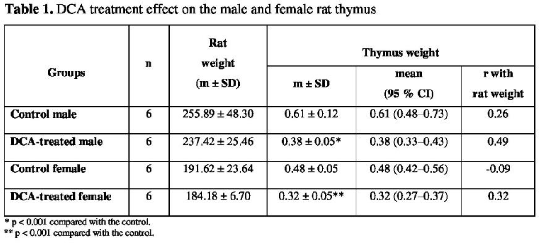
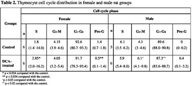
Results: The DCA treatment effect on thymus of both genders is presented in Table 1. In gonad-intact rats of both genders, the thymus weight of the control was higher than in rats treated with DCA (p < 0.001). Flow cytometry showed (Table 2) that DCA treatment increased the percentage of thymocytes in the G2-M phase (p < 0.03) and reduced in G1-G0 (p < 0.02) in males, and in the females the percentage of cells increased in the S phase (p < 0.024) and reduced in the pre-G phase (p < 0.034) as compared with the controls.


Conclusion: The thymocyte cell cycle analysis showed the gender-related distinction of the DCA effect which may be important when analysing its pharmacological efficacy in further preclinical investigations.
References:
1. Zhang SL et al. (2016). J Med Chem 59: 3562-3568.
2. Stander XX et al. (2015). Cell Physiol Biochem 35: 1499-1526.

