

| 014P Nottingham, UK 7th Focused Meeting on Cell Signalling |
Receptor phosphorylation is key for agonist-induced internalisation of M1 muscarinic acetylcholine receptors
Introduction: M1 muscarinic acetylcholine receptor (M1 mAChR) agonists and positive allosteric modulators improve cognition and enhance cholinergic signalling in Alzheimer’s disease models1-3. However, these ligands can show a wide range of adverse effect liabilities associated with on-target overstimulation of the receptor4. Recently, our lab have demonstrated that transgenic mice expressing a phoshorylation-deficient mutant of the M1 mAChR, are more sensitive to adverse responses, such as seizures, in response to administration of M1-selective agonists. We hypothesise that M1-agonists stimulating increased phosphorylation and internalisation may be less likely to induce receptor overstimulation and on-target adverse effects. In this study, we aim to understand the role of phosphorylation in agonist-induced internalisation specifically.
Method: Cell monolayers of CHO cells expressing wild-type (WT) M1 muscarinic acetylcholine receptors (mAChRs) or the phosphorylation-deficient mutant (PD) where all serine residues in the third intracellular loop and C-terminal tail are mutated to alanine, were stimulated for 0, 5, 15 and 30, 60 and 240 minutes with the muscarinic agonist carbachol (CCh; 100 μM). CCh-treated cells were transferred on ice, washed with ice-cold Krebs-Henseleit buffer and radiolabelled with saturating concentration of [3H]-NMS (4 nM) overnight at 4°C to quantify cell surface expression. All data are expressed as means ± S.E.M. of three separate experiments (n=3) and were normalised as percentage of untreated cells for each assay.
Results: In M1 mAChR WT cells, four hours of CCh stimulation caused a reduction in cell surface receptors to to 58.4% ±2.2% compared to untreated cells, with most of the receptors (29.9% ±7.1%) internalising within the first hour (Fig. 1). In contrast, agonist treatment of cells expressing PD mutant of M1 AChR caused no change in levels of surface receptors within the first hour of stimulation and 8.4% ±3.4% receptor internalisation after 4 hours , which may be associated with receptor desensitisation or constitutive internalisation rather than agonist-induced internalisation.
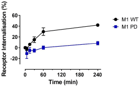
Conclusions: Agonist-induced internalisation of M1 mAChRs is mediated by receptor phosphorylation and it was prevented when receptor phosphorylation sites were removed. Receptor phosphorylation is crucial for agonist-induced internalisation and is likely contributing to an increased adverse effect liability of M1-selective agonists in the M1 PD mice.
Reference:
1 Bodick, N. C. et al. (1997). Archives of Neurology 54, 465-473.
2 Puri, V., et al. (2015). Behavioural Brain Research 287, 96-99.
3 Bradley, S. J. et al. (2017). J Clin Invest. 127, 487-499.
4 Rook, J. M. et al. (2017). ACS Chemical Neuroscience 8, 866-883.