

| 045P Nottingham, UK 7th Focused Meeting on Cell Signalling |
Analysis of inverse agonist activity of ticagrelor at the P2Y12 receptor
Introduction: Acute coronary syndrome (ACS) is often caused by atherosclerotic plaque rupture, resulting in activation and aggregation of platelets. Activated platelets release ADP stimulating the P2Y12 receptor (P2Y12R), responsible for amplification of the platelet response. Current ACS treatment includes the P2Y12R “blocker” ticagrelor, which has shown improved clinical efficacy over other P2Y12R antagonists including clopidogrel. We have previously demonstrated inverse agonist properties of ticagrelor in platelets (1). The aim of this study is to further investigate the possible inverse agonism of ticagrelor, and compare its mechanism of action against other P2Y12 inhibitors, AR-C66906 and R-138727.
Method: Effects of ticagrelor on G protein activation of P2Y12R were monitored with bioluminescence resonance energy transfer (BRET2), as previously described (2). HEK293 cells were transiently transfected with FLAG-tagged P2Y12R and 48 h later seeded into a 96 well plate at 1×10^5 /plate and incubated with ticagrelor (10uM) or vehicle (0.1 % DMSO) for 5 minutes. To assess P2Y12R surface levels, flow cytometry was performed on washed platelets as previously described (3). Platelets were incubated for 1 h with vehicle (1% DMSO or HEPES-Tyrode’s buffer) or ticagrelor, AR-C66096 or R-138727 (10 μM), before fixation and labelling. Data is plotted as mean BRET ratio or geometric mean fluorescence intensity (MFI) ± SEM, and analysed via a two-way ANOVA, with Bonferroni corrections.
Results: P2Y12R-transfected cells showed an attenuated BRET ratio compared to pcDNA 3.1-transfected cells, which was partially reversed by ticagrelor (10 μM) (table 1). Flow cytometry assays indicated an increased MFI in response to ticagrelor (10 μM) compared to vehicle. AR-C66096 and R-138727 did not affect MFI (figure 1).
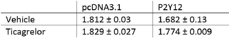
Table 1. BRET analysis of P2Y12R activity in pcDNA3.1- and P2Y12R-transfected HEK293 cells in response to 10 μM ticagrelor (n = 3).
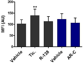
Figure 1. Flow cytometry of P2Y12R receptor levels in washed platelets (**p<0.01 ticagrelor vs vehicle, n = 7).
Conclusion: BRET analysis indicates ticagrelor inhibits the constitutive activity of P2Y12R. Additionally, ticagrelor, but not AR-C66906 or R-138727, increase P2Y12R surface expression in platelets. This data suggests inverse agonist properties of ticagrelor, which may contribute to increased clinical efficacy.
References:
(1) Aungraheeta R et al. (2016). Blood 128(23): 2171-2727.
(2) Pfleger KDG. (2006) Nature Protocols 11: 337-345.
(3) Mundell SJ et al. (2018). J Thromb Haemost 16: 44-53.