

| 1155 Virtual Meeting BPS & ELRIG UK joint meeting: Translating Ideas into Therapies |
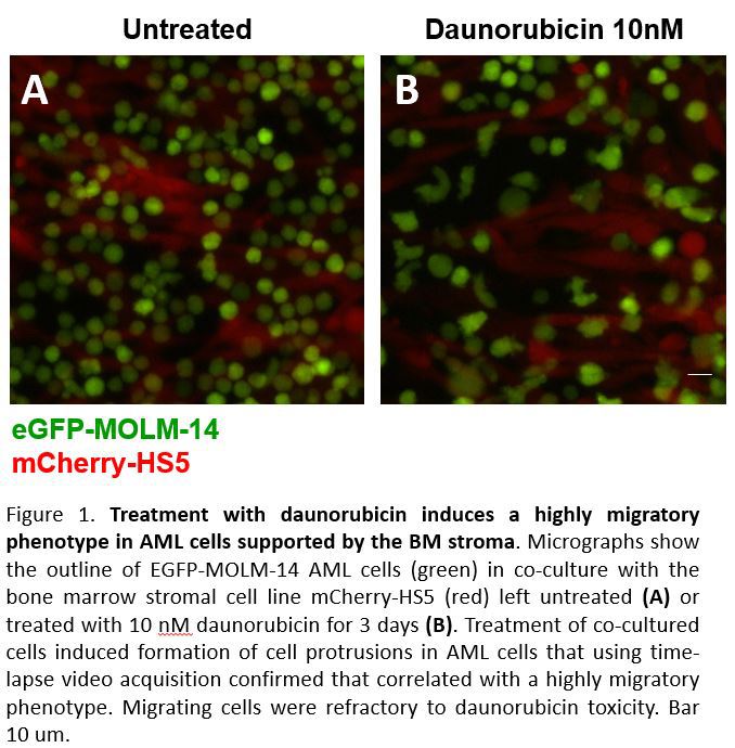
Therapeutic drugs promote differentiation of AML leukemic blasts into a migratory and chemo-resistant phenotype facilitated by the bone marrow microenvironment.
Introduction/Background & aims Although current therapies induce high rates of remission in AML, there is still an unmet need for new therapeutic strategies as the majority of patients will relapse and become refractory. The adhesive interactions between AML leukemic stem cells and the bone marrow (BM) microenvironment are crucial during AML development and resistance to drug treatments1. Therefore, improving experimental approaches for AML studies as well as drug screening methods require the development of in vitro co-culture models that closely recreate the protective BM setting. We have created a fluorescence-based in vitro model scalable to high throughput that reproduces the chemo-resistance mediated by the BM stroma. Using our model, we verified that the therapeutic effect of both daunorubicin and cytarabine (the major clinical drugs currently used against AML) was inhibited by co-culture with BM stromal cells. Chemo-resistant AML cells attached on the BM stroma and became highly migratory. We investigated further this unexpected phenotype.
Method/Summary of work We generated a panel of clonal Human AML cell lines stably expressing eGFP that were selected for showing linear correlations between the number of seeded cells and the fluorescent signal with equivalent sensitivity to the MTT assay. The cell lines include Kasumi-1, MV4-11, THP-1, KG-1, U937, MOLM14 and HL-60, which bear various genetic alterations representative of AML. As BM stromal cells we used the fibroblastic HS5 cell line transduced with the red fluorescent protein mCherry that we have previously generated, validated and used in former studies2. eGFP-AML cell lines and mCherry-HS5 cells were co-cultured and left untreated or treated with the corresponding reagents. Changes in eGFP/mCherry signal was used as a readout for changes in AML/BM stromal cell numbers to calculate their proliferation index and allowed us to distinguish the cell types when filmed using time lapse microscopy. Cytokine and phosphoproteomic assays were performed as well as cell cycle analysis by flow cytometry and qPCR.
Results/Discussion AML chemo-resistant cells displayed a significant increase in cell size, cytoplasmic complexity and motility in the presence of cytoprotective BM stromal cells while simultaneously upregulating CD163 and CD197 (M1 and M2 macrophage maturation markers, respectively). These unusually differentiated AML cells adopted a polarised migratory morphology with well-formed vimentin filaments as well as organised F-actin in lamellipodia and formation of robust actin-based adhesive structures called podosomes characteristic of normal myeloid cells macrophages and dentritic cells. The increased migration of resistant AML cells also correlated with upregulation of the epithelial mesenchymal transition (EMT)-related genes previously correlated with aggressive solid tumours and more recently with poor prognosis in AML patients carrying MLL translocations. The genes include: ZEB1, EIF4A3, LPS1, MAP7, STK17B, TRPS1 and VIM. Surviving AML cells were also restrained in the S-phase of the cell cycle and showed higher levels of the stem cell marker CD44. The BM stromal cell secretome directly induced the migratory and abnormally differentiated AML phenotype and was enriched in myeloid differentiation and chemotactic factors including GM-CSF, MCP-1, SDF1alpha, IL-1ra, GRO-alpha and IP-10.
Taking advantage of the high throughput capacity of our experimental platform, we performed phosphoproteomic and drug screening studies that converged in identifying JAK1/2 kinases as critical mediators for BM microenvironment-mediated chemo-resistance to daunoribicin. The JAK1/2 inhibitors Ruxolitinib and Baricitinib sensitised AML cells to daunorubicin in the presence of BM stromal cells and inhibited by 70% daunorubicin-induced increased migration.
Conclusion(s) Taken together, our results indicate that daunorubicin and cytarabine induce secretion of myeloid differentiating and immunosuppresive cytokines in the tumour microenvironment leading to an abnormal macrophage maturation phenotype in AML blasts that results in resistance to drug treatment and this process requires activation of JAK1/2 kinases. We propose that using JAK inhibitors in combination with daunorubicin may prevent cell adhesion mediated drug resistance in AML leading to improved therapies for this currently incurable disease.
Reference(s)
1. Chen, P. et al. Bone marrow stromal cells protect acute myeloid leukemia cells from anti-CD44 therapy partly through regulating PI3K/Akt-p27 axis. Mol.Carcinog. (2014).
2. Ramasamy, K. et al. Fluorescence-based experimental model to evaluate the concomitant effect of drugs on the tumour microenvironment and cancer cells. British journal of haematology 157, 564-579, doi:10.1111/j.1365-2141.2012.09103.x (2012).