| pA2 online © Copyright 2004 The British Pharmacological Society |
027P
University of Buckingham 3th Focused Meeting April 2004 |
|
Immunocytochemical
localisation of the novel obesity peptide, neuropeptide W and its
receptors GPR7 and GPR8 to human cardiovascular and renal tissues |
|
We have
previously described the localisation of the novel obesity peptide, neuropeptide
W (NPW), and its receptor GPR7, in rat brain (Singh et al., 2004).
In humans, NPW also binds to GPR8, not yet detected in rats and mice (Shimomura
et al., 2002). Using antisera to the peptide and its target receptors,
the distribution of this novel transmitter system was determined in human
peripheral tissues.
Acetone fixed sections (30µm) of fresh frozen human tissue (left
ventricle, lung, kidney and left internal mammary artery) were blocked
for 1 hr with 5% swine serum. Sections were incubated with 1:200 dilution
of rabbit anti-NPW antisera (Phoenix Pharmaceuticals Inc., CA, U.S.A)
for 24 hrs at 4oC. Binding of the primary
antisera to the peptide was visualised using the peroxidase anti-peroxidase
method. Tissues in which NPW-like immunoreactivity (LI) was detected were
then examined for receptor distribution with either the human GPR7(178-194)
or GPR8(25-45) antisera (Lifespan
Biosciences Inc.) at 1:50 dilution.
In human kidney, intense NPW-LI (Figure 1A) was visualised in the glomeruli
of the renal cortex while faint staining was also observed within small
diameter renal vessels. Specific staining was not detected in renal tubules
or the medullary rays. Furthermore, NPW-LI was visualised in the endocardial
endothelium cells as well as in the endothelium of both intramyocardial
vessels and the left internal mammary artery. Immunoreactive NPW staining
was not detected within any cell type in human lung.
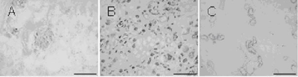
Figure
1: Presence of NPW-LI (A) in the glomeruli of the renal cortex and GPR7-LI
(B) and GPR8-LI (C) detected in renal tubules of the renal medulla. Scale
bar = 200 µm.
As intense NPW-LI was observed in human kidney, sections of the renal
system were examined for NPW receptor staining. In the renal medulla,
intense and abundant GPR7-LI (Figure 1B) was detected within the renal
tubules but not in the medullary rays. GPR8-LI (Figure 1C) was also observed
in renal tubules of the medulla but was discretely distributed in comparison
to GPR7-LI. Immunoreactivity for both GPR7 and GPR8 was not detected in
the renal cortex or renal vessels.
We believe this to be the first evidence of identification of NPW and
its receptor in human tissues. The presence of receptor and peptide in
the kidney suggests a potential role for this novel transmitter in renal
function. Furthermore, the observed NPW-LI in the endothelium of the left
internal mammary artery and presence of NPW in the glomeruli might imply
NPW to be a circulating peptide.
Shimomura et al., (2002). J Biol Chem; 277, 35826-35832
Singh et al., (2004).
http://www.pa2online.org/Vol1Issue3abst016P.html